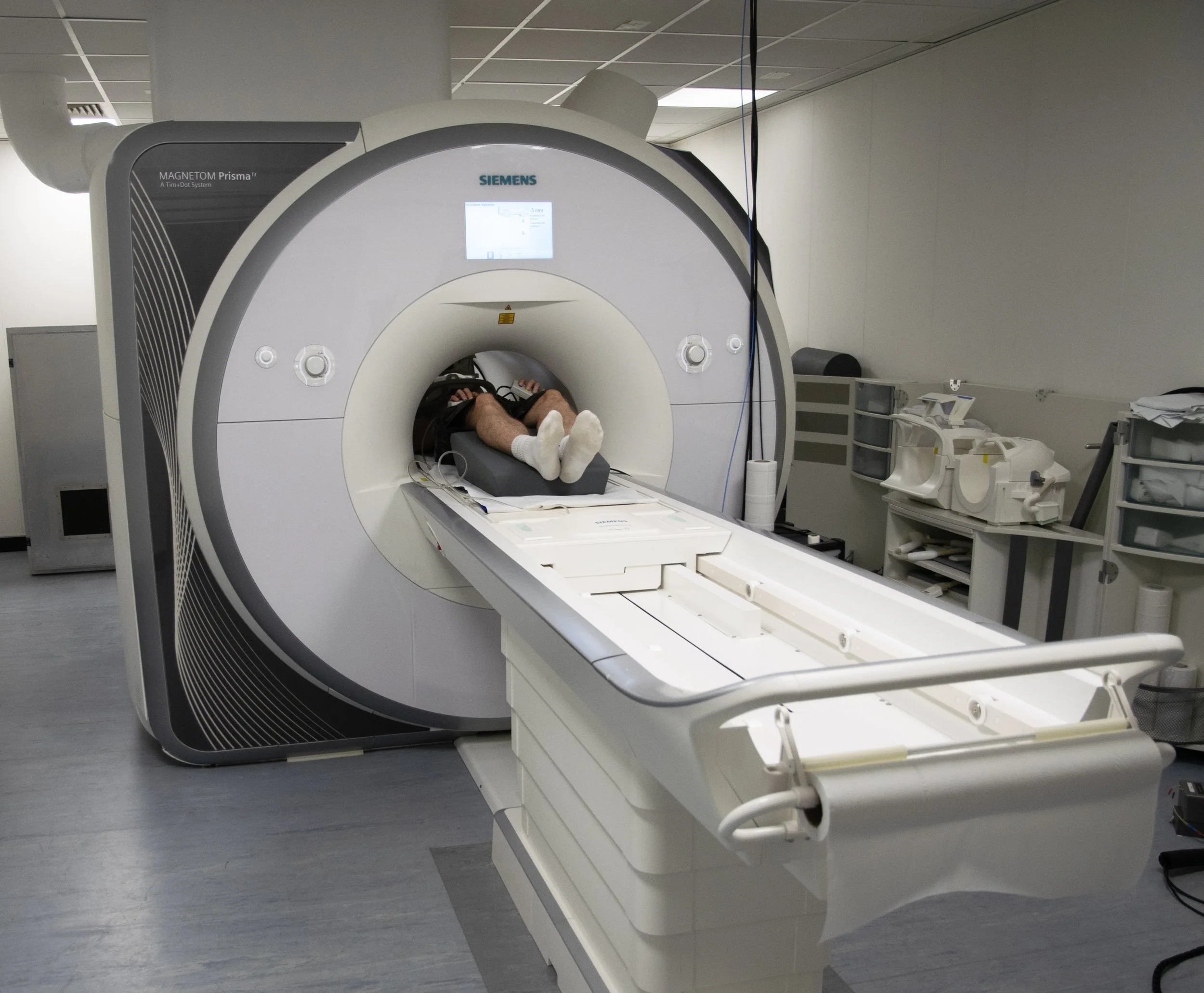
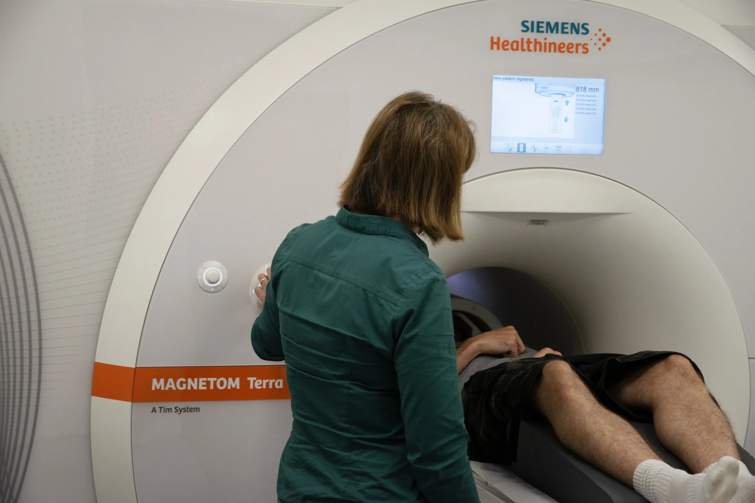
3T MRI Technology
What is 3T Technology? FAQs
Magnetic Resonance Imaging (MRI) is a type of scan that uses strong magnetic fields and radio waves to produce highly detailed images of any part of the body, such as the brain. MRI is extensively used in the medical field for diagnosis and monitoring of disease and injury as well as research purposes. Our scans are strictly for research purposes, and are not diagnostic.
We mostly use 2 types of MRI scans:
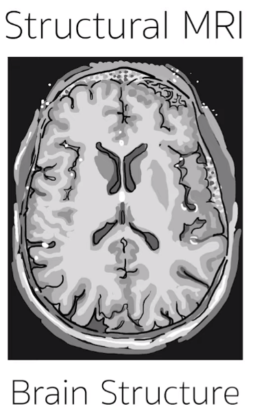
Structural – for seeing detailed images of structures within the brain.
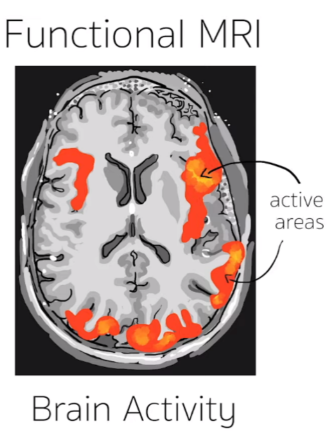
Functional – for seeing which parts of the brain are active when you perform certain tasks. This is done by detecting changes in the oxygen levels in the blood.
-
MRI scans take place in a strong magnetic field. Therefore, no metallic items can be taken into the scanner since they will get attracted by the magnet and may get heated up by the radio waves. For the same reason, people with certain metallic implants cannot be scanned. Such metal items include any of the following: Cardiac pacemakers, cochlear implants, metallic aneurysm clips, metallic fragments in the eye and certain types of bio -mechanical implants. If in doubt, please contact your researcher. Although there is no evidence to suggest that MRI is harmful to unborn babies, as a precaution, the Department of Health advises against scanning pregnant women unless there is a clinical benefit. We do not test for pregnancy as routine so if you think you may be pregnant you should not take part in this study.
-
MRI is a painless and safe technique to obtain detailed pictures of the brain. Unlike X-rays, MRI does not involve exposing the body to radiation, but uses magnets instead. MRI scanning is a safe technique, as long as you do not have any metal on your person or in your body. Before being invited to the Centre, a researcher will check that you are suitable for scanning by asking a number of safety questions.
-
You will be asked to leave ANY metallic objects you may be carrying. Clothing should be loose and comfortable (not sports or ‘technical’ fabrics) with minimal metal/metallic items. Any bras must be removed. Medical scrubs can be provided. Glasses cannot be worn inside the scanner; however we do provide MRI safe glasses. Our available prescriptions range from -6 to +6. You will be asked to remove any make up prior to going into the scanner as certain types can interfere with the quality of the data. You must complete a screening questionnaire and a consent form, as well as provide details of your UK based GP.
-
One of our imaging team members will guide you into the MRI room safely and will ask you to lie down on a comfortable bed. Cushions or supports for your knees and back may be used to make you more comfortable. Blankets are available if needed. The scanning process generates intermittent loud buzzing/tapping noises therefore you will be given earplugs to wear or special headphones to wear during the scan. A special device, known as a head coil, will be placed over your head. This receives the signals that are used to create the images. Once you feel comfortable and happy to proceed, you will be moved inside the scanner and will be asked to keep very still while the scan takes place as movement can interfere with data quality. Our imaging team members will assist you throughout the procedure and you will be able to talk with them through an intercom at any time. If you feel uncomfortable you can call them just by pressing a buzzer which will be in your hand at all times during the scan. Research scan sessions tend to be longer than standard clinical scans and you will be given instructions about tasks and scan duration by your researcher.
-
After the session has finished we will remove you from the scanner, and the researcher will accompany you to collect your belongings. Your GP will not be notified of your participation in this research.
-
Rarely, some individuals may experience a tingling sensation in the peripheral parts of the body. This is an infrequent side effect of MRI scanning. If this occurs, you will be trained in advance to press the squeeze bulb, and the scan will immediately be stopped.
-
Yes, it is safe to have multiple MRI scans because the magnetic fields used to produce the images are “non-ionizing” meaning that they do not cause harm to your body. There are no known negative consequences of imaging when the scanning is conducted appropriately.
-
Depending on the study that you are participating in, it may be possible for you to receive a picture of your brain. Please ask the Researcher for the study that you are taking part in.
-
If you have something implanted in your body, e.g. as a result of a surgery that you underwent, it is possible that the implant can heat up during scanning. Many implants have been evaluated by the manufacturers who provide us with information about how to safely scan people with them. However, if this information is not available then we may not be able to go ahead with scanning because we have to be very careful about safety when the scan is not required for your explicit clinical benefit.
-
Should the MRI scan unexpectedly reveal any clinically relevant abnormality, we would like your permission to notify your GP. However, please be aware that this process will only happen where there is a clear abnormality in your scan and you should not participate in a study with the purpose of obtaining a diagnosis.
What is the difference between 3T and 7T MRI?
7T MRI simply uses a stronger magnet to take images of the brain compared to 3T MRI scanners. The stronger magnet produces a bigger signal which means that we can look at even small structures in the brain. Magnetic susceptibility is a property of all matter and the impact of the tissue’s susceptibility is much bigger when in a stronger magnetic field. This means that 7T MRI can also produce images with much better contrast between structures, particularly in the case of functional MRI which relies on susceptibility changes that occur in our blood when we engage in activities, e.g. seeing, hearing, or remembering. With 7T MRI we can actually look at signals from different layers of the cortex. This is really important for understanding the mechanisms behind how we do things.
Example benefits of 7T relative to 3T:

What can 3T MRI Technology be used for?
MRI is a really flexible technology and can be used to explore many different aspects of the human brain. A few examples are highlighted below:
We can explore the functional organisation of the brain using a technique called functional MRI to better understand how the brain supports particular behaviours and what goes wrong in psychiatric or neurological disorders.
We can obtain images that tell us about the brain’s structural organisation. For example, we can tailor our measurements to be sensitive to particular biological quantities such as myelination, iron or water content.
We can characterise the vascularity of the brain to understand how oxygen is delivered to different regions of the brain.
By examining how water diffuses on the microscale we can map the trajectory of different fibre pathways in the white matter that connects different regions of our brains.
To find out about studies which involve MRI brain scanning, please see the News section of our website.